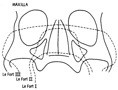

In a Le Fort I fracture, the tooth-bearing part of the maxilla becomes separated from the rest of the maxilla by fractures through the medial and lateral walls of the maxillary sinuses, and by a fracture through the lower part of the nasal septum.
The Le Fort II fracture is more extensive and the separated fragment is pyramidal. The apex of the pyramid is the lower part of the nasal bones, and the fracture lines run inferiorly and laterally through the medial and inferior walls of the orbits, and then through the anterior and posterolateral wall of the maxillary sinuses. The nasal septum is fractured at a variable level.
In the Le Fort III fracture, there is complete craniofacial dysjunction. The fracture line runs through the nasal bones as in the Le Fort II fracture, and then posteriorly and laterally through the medial and lateral walls of the orbits, and through the zygomatic arches. The nasal septum is fractured superiorly.
Leave a Reply